Bdsm

BDSM Research Widget

BDSM Community Research Widget

Anonymous • Ethical • 5–8 min total

Interview Question Bank

Pick 3–5 to ask per person. Tap to select.

Ice-Breakers

Surface-Level

Deeper (with rapport)

Copied!

DASS-21 (Past Week)

0 = Never • 1 = Sometimes • 2 = Often • 3 = Almost Always

Your DASS-21 Scores (x2 scaled)

Depression:

Anxiety:

Stress:

These are for research only. Not a diagnosis.

Optional Demographics

Ready to Submit?

Your data is 100% anonymous. No IP, no names.

Debrief

Thank you! This research helps reduce stigma around mental health in kink communities.

Support: Kink-Aware Professionals • 800-656-HOPE

Tuesday, July 16, 2024

Methamphetamine's Toll on the Brain: A Deep Dive into Cellular Destruction

Methamphetamine (METH), a highly addictive stimulant, is wreaking havoc on brains worldwide. This potent drug not only alters mood and behavior but also inflicts severe damage at the cellular level.

How Methamphetamine Attacks the Brain

At the heart of METH's destructive power lies its ability to hijack the brain's reward system. By flooding the brain with dopamine, METH induces a euphoric high that quickly becomes addictive. However, the long-term consequences are devastating (D'Brant et al, 2019):

  1. Mitochondrial Mayhem: METH molecules infiltrate mitochondria, the cell's powerhouses, disrupting energy production and leading to cell death.
  2. Glial Cell Breakdown: Glial cells, essential for brain function, are also victims of METH's assault, contributing to overall brain damage.
  3. Apoptosis: Cellular Suicide: METH triggers programmed cell death, or apoptosis, accelerating brain tissue degeneration.

Unraveling the Mystery with Advanced Imaging

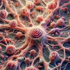
To understand the full extent of METH's devastation, scientists are employing cutting-edge imaging techniques (D'Brant et al, 2019):

  1. 3D Tomographic Imaging: This technology creates detailed 3D images of cells without harmful dyes, allowing researchers to observe structural changes in real time.
  2. Digital Holographic Microscopy (DHM): By measuring subtle changes in light, DHM provides precise information about cell volume and shape, helping to track the progression of cell death.
  3. Raman Spectroscopy: This technique analyzes the molecular composition of cells, revealing chemical changes associated with METH-induced damage.

Researchers employed advanced imaging techniques to observe the behavior of glial cells exposed to METH. By comparing these cells to those exposed to a known cell-killing drug, doxorubicin, they were able to identify specific changes caused by METH (D'Brant et al, 2019):


  1. Cell Shrinkage: Glial cells exposed to METH experienced a significant decrease in size, a hallmark of cell death.
  2. Mitochondrial Damage: METH also caused a reduction in the size of mitochondria, the cell's energy powerhouses.
  3. Rapid Progression: The effects of METH on cell volume were observed within just 40 minutes of exposure.
  4. Chemical Changes: Raman spectroscopy revealed alterations in the chemical composition of cells exposed to METH, indicating broader cellular damage beyond cell death.  

  Conclusion

Methamphetamine's destructive power lies in its ability to disrupt brain chemistry and induce cellular damage. By overwhelming the brain with dopamine and attacking vital cell components like mitochondria, METH triggers a cascade of events leading to cell death. Advanced imaging techniques have revealed this cellular devastation's rapid and severe nature. Understanding these mechanisms is crucial for developing effective prevention and treatment strategies for methamphetamine addiction.      


References

  1.  D’Brant, L. Y., Desta, H., Khoo, T. C., Sharikova, A. V., Mahajan, S. D., & Khmaladze, A. (2019).                    Methamphetamine-induced apoptosis in glial cells examined under marker-free imaging modalities.             Journal of Biomedical Optics, 24(4), 046503. https://doi.org/10.1117/1.JBO.24.4.046503


Monday, July 15, 2024

Spotting Alzheimer's Early: Promising New Tools on the Horizo

Alzheimer's disease (AD), a progressive brain disorder leading to memory loss and cognitive decline, affects millions worldwide. Early detection is key for managing symptoms and planning for the future. However current diagnostic methods can be expensive and invasive, involving procedures like lumbar punctures and PET scans.  Moreno and colleagues (2024) inform us that new research is exploring exciting possibilities for detecting AD earlier and with less hassle. Their research points towards two promising avenues: EEG (electroencephalography) and the gut microbiome.

Shining a Light on Brain Activity: EEG

EEG is a non-invasive technique that measures electrical activity in the brain using electrodes placed on the scalp. It's similar to the technology used in sleep studies. Moreno and colleagues (2024) write that researchers are investigating whether EEG patterns can differentiate between healthy individuals and those with AD:

  • The initial findings look promising. Studies suggest that people with AD exhibit a distinct EEG signature compared to healthy controls. This signature involves a slowing of overall brain activity, with a decrease in the "fast" brain waves and an increase in the "slow" ones.

The Gut Connection: Your Microbiome and Your Brain

The trillions of microbes living in your gut, collectively called the gut microbiome, play a crucial role in overall health. Recent research suggests a fascinating link between the gut microbiome and brain health.

Gut Microbiome and AD: A Shifting Landscape

Moreno and colleagues (2024) tell us more about this shifting landscape:

  1. The gut microbiome plays a significant role in human health, and its diversity naturally declines with age. 
  2. Interestingly, research has shown that individuals with AD have a distinct gut microbiome composition compared to healthy people. 
  3. This imbalance, called gut dysbiosis, involves a decrease in beneficial bacteria and an increase in potentially harmful ones. This dysbiosis is linked to chronic inflammation, a risk factor for AD.


How the Gut Talks to the Brain: The Gut-Brain Axis

So how does what happens in your gut impact your brain? It all comes down to communication. The gut and brain are connected through a complex network called the gut-brain axis. When gut dysbiosis occurs, it can lead to increased gut permeability, often referred to as "leaky gut." This allows harmful substances to enter the bloodstream and potentially reach the brain, potentially worsening AD (Moreno et al, 2024).

Microbiome Metabolites: Friend or Foe?

The bacteria in our gut not only interact with our body but also produce chemicals called metabolites that can influence the nervous system. Short-chain fatty acids (SCFAs) are a type of beneficial metabolite produced by gut bacteria. They help reduce inflammation and may even improve cognitive function.  On the other hand, elevated levels of another metabolite called Trimethylamine N-oxide (TMAO) have been linked to cognitive decline and AD pathology. (Moreno et al, 2024)

Earlier Detection, Better Outcomes

These new methods, EEG and gut microbiome analysis, offer a potentially revolutionary approach to AD detection. They are non-invasive, potentially less expensive, and could pave the way for earlier diagnosis. This earlier detection would allow for earlier intervention and treatment, potentially improving patient outcomes and quality of life.

Looking Ahead: The Road to Better Diagnosis

While the research on EEG and the gut microbiome in AD diag

nosis is promising, it's still in its early stages. More studies are needed to validate these findings and determine how best to incorporate these tools into clinical practice.

However, the potential for earlier, less-invasive AD detection is a significant step forward.  This research offers a glimmer of hope for a future where Alzheimer's can be identified and managed before it significantly impacts a person's life


References

  •  Moreno, Diego A. & Ramos-Molina, Bruno & Andjelkovic, Anuska & Ruiz-Alcaraz, Antonio & Krothapalli, Mahathi & Buddendorff, Lauren & Yadav, Hariom & Schilaty, Nathan & Jain, Shalini. (2024). International Journal of Molecular Sciences Review From Gut Microbiota to Brain Waves: The Potential of the Microbiome and EEG as Biomarkers for Cognitive Impairment. International Journal of Molecular Sciences. 25. 6678. 10.3390/ijms25126678. 

Combating Methamphetamine's Devastating Effects: A New Hope for Cognitive Recovery

Methamphetamine (METH) is a highly addictive stimulant that wreaks havoc on the brain and behavior. This highly prevalent drug not only creates intense cravings but also leads to a significant decline in cognitive function over time. With current treatments focusing on managing addiction itself, a new approach targeting the cognitive damage caused by METH offers a glimmer of hope.

METH's Deleterious Impact on the Brain

Chronic METH use disrupts the brain's reward system, specifically areas like the Ventral Tegmental Area (VTA) and Nucleus Accumbens (NAc). This disruption leads to intense cravings and compulsive drug-seeking behavior, making it incredibly difficult to quit. Additionally, METH significantly impairs cognitive abilities, impacting memory, learning, and decision-making. This decline in cognitive function poses a major challenge for individuals struggling with METH addiction.

Treatment Challenges and a Promising New Direction

Currently, there's no single magic bullet for METH use disorder. However, research suggests that addressing cognitive deficits could be a valuable complementary approach. Studies using Memantine, Berberine, and Melatonin in animal models have shown promise in improving cognitive function after METH exposure.

Paeoniflorin (PF): A Natural Light on the Horizon

Paeoniflorin (PF) is a natural compound extracted from the Paeonia lactiflora plant. This compound boasts a range of therapeutic properties, including anti-inflammatory, antioxidant, and neuroprotective effects. These properties have made PF a potential candidate for treating neurodegenerative diseases like Alzheimer's and Parkinson's. Notably, studies show PF's success in reducing cognitive decline and inflammation in animal models of these diseases. Furthermore, PF seems to improve spatial learning and memory function, crucial aspects of overall cognitive well-being.

A New Study Tackles METH-Induced Cognitive Decline

A groundbreaking new study is investigating the potential of PF to counteract the cognitive impairment caused by METH in mice. This study utilizes various tests, including new location recognition (NLR), new object recognition (NOR), and Y-maze tasks, to assess the cognitive function of mice treated with both PF and METH. Additionally, the study examines how PF might influence reward-seeking behavior induced by METH. 

To delve deeper into the potential mechanisms of PF, researchers will analyze changes within brain regions like the VTA, NAc, and Hippocampus. These regions are crucial for memory and synaptic function, and the study will investigate changes in protein levels associated with these functions (PSD-95 and synaptophysin) to understand how PF might be working.

Overall, this exploration of PF as a potential treatment for the cognitive deficits linked to METH use disorder is a compelling development. By investigating both the cognitive and behavioral effects of PF, this study offers valuable insights into a promising new therapeutic avenue for individuals struggling with the long-term consequences of METH addiction.

Reducing Cravings and Protecting the Brain

The research showed that PF successfully reduced two key aspects of METH addiction in mice (Gong et al, 2024):

  1. Expression and reinstatement of conditioned place preference (CPP): CPP is a behavioral test used to measure an animal's association of a place with a rewarding experience. The study suggests PF helps prevent the development of cravings associated with METH use.
  2. Synaptic protein levels: METH can increase levels of specific proteins linked to addiction in the brain. PF appears to counteract this effect, potentially protecting brain cells from damage caused by METH.

What This Means

This research is a significant step forward. While the study was conducted on mice, it lays the groundwork for further investigation into PF's potential as a treatment for METH addiction in humans.

Important Note:

It's crucial to remember that this is preliminary research. More studies are needed to confirm these findings and determine PF's safety and efficacy in humans.

The Future of Addiction Treatment

This research offers a promising lead in the fight against METH addiction. PF's potential to reduce cravings and protect brain cells is a significant development. As research progresses, we may see PF emerge as a valuable tool for helping people overcome METH addiction and reclaim their lives.


References

  1. Gong, Xinshuang & Yang, Xiangdong & Yu, Zhaoying & Lin, Shujun & Zou, Zhiting & Qian, Liyin & Ruan, Yuer & Si, Zizhen & Zhou, Yi & Li, Yu. (2024). The effect of paeoniflorin on the rewarding effect of METH and the associated cognitive impairment in mice. 10.21203/rs.3.rs-4430457/v1. 

Featured Blog Post

Breaking the Cycle: How Meth and GHB Mess with Your Brain's Wiring

Hey there, if you're reading this, you're probably knee-deep in that exhausting loop of highs, crashes, and compulsions that come wi...