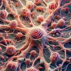
How Methamphetamine Attacks the Brain
At the heart of METH's destructive power lies its ability to hijack the brain's reward system. By flooding the brain with dopamine, METH induces a euphoric high that quickly becomes addictive. However, the long-term consequences are devastating (D'Brant et al, 2019):
- Mitochondrial Mayhem: METH molecules infiltrate mitochondria, the cell's powerhouses, disrupting energy production and leading to cell death.
- Glial Cell Breakdown: Glial cells, essential for brain function, are also victims of METH's assault, contributing to overall brain damage.
- Apoptosis: Cellular Suicide: METH triggers programmed cell death, or apoptosis, accelerating brain tissue degeneration.
Unraveling the Mystery with Advanced Imaging
To understand the full extent of METH's devastation, scientists are employing cutting-edge imaging techniques (D'Brant et al, 2019):
- 3D Tomographic Imaging: This technology creates detailed 3D images of cells without harmful dyes, allowing researchers to observe structural changes in real time.
- Digital Holographic Microscopy (DHM): By measuring subtle changes in light, DHM provides precise information about cell volume and shape, helping to track the progression of cell death.
- Raman Spectroscopy: This technique analyzes the molecular composition of cells, revealing chemical changes associated with METH-induced damage.
Researchers employed advanced imaging techniques to observe the behavior of glial cells exposed to METH. By comparing these cells to those exposed to a known cell-killing drug, doxorubicin, they were able to identify specific changes caused by METH (D'Brant et al, 2019):
- Cell Shrinkage: Glial cells exposed to METH experienced a significant decrease in size, a hallmark of cell death.
- Mitochondrial Damage: METH also caused a reduction in the size of mitochondria, the cell's energy powerhouses.
- Rapid Progression: The effects of METH on cell volume were observed within just 40 minutes of exposure.
- Chemical Changes: Raman spectroscopy revealed alterations in the chemical composition of cells exposed to METH, indicating broader cellular damage beyond cell death.
Conclusion
Methamphetamine's destructive power lies in its ability to disrupt brain chemistry and induce cellular damage. By overwhelming the brain with dopamine and attacking vital cell components like mitochondria, METH triggers a cascade of events leading to cell death. Advanced imaging techniques have revealed this cellular devastation's rapid and severe nature. Understanding these mechanisms is crucial for developing effective prevention and treatment strategies for methamphetamine addiction.
References
- D’Brant, L. Y., Desta, H., Khoo, T. C., Sharikova, A. V., Mahajan, S. D., & Khmaladze, A. (2019). Methamphetamine-induced apoptosis in glial cells examined under marker-free imaging modalities. Journal of Biomedical Optics, 24(4), 046503. https://doi.org/10.1117/1.JBO.24.4.046503


No comments:
Post a Comment